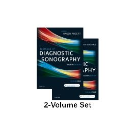
Test Bank for Textbook of Diagnostic Sonography 8th Edition by Hagen Ansert
$35.00 Original price was: $35.00.$26.50Current price is: $26.50.
Test Bank for Textbook of Diagnostic Sonography 8th Edition by Hagen Ansert
This is completed downloadable of Test Bank for Textbook of Diagnostic Sonography 8th Edition by Hagen Ansert

Product Details:
- ISBN-10 : 0323353754
- ISBN-13 : 978-0323353755
- Author: Hagen Ansert
Updated to reflect the newest curriculum standards, Textbook of Diagnostic Sonography, 8th Edition provides you with the pertinent information needed for passing the boards. This highly respected text enhances your understanding of general/abdominal and obstetric/gynecologic sonography, the two primary divisions of sonography, as well as vascular sonography and echocardiography. Each chapter covers patient history; normal anatomy, including cross-sectional anatomy; sonography techniques; pathology; and related laboratory findings. And more than 3,100 images and anatomy drawings guide you in recognizing normal anatomy and abnormal pathology.
Table of Content:
- PART I. Foundations of Sonography
- 1. Foundations of clinical sonography
- The role of the sonographer
- Historical overview of sound theory and medical ultrasound
- Introduction to basic ultrasound principles
- Bibliography
- 2. Essentials of patient care for the sonographer
- A sonographer’s obligations
- Basic patient care
- Patients on strict bed rest
- Patients with tubes and tubing
- Patient transfer techniques
- Infection control
- Isolation techniques
- Emergency medical situations
- Professional attitudes
- Assisting patients with special needs
- Evaluating patient reactions to illness
- Patient rights
- Bibliography
- 3. Ergonomics and musculoskeletal issues in sonography
- History of ergonomics
- Injury data in sonography
- Industry awareness and changes
- Work practice changes
- Economics of ergonomics
- References
- 4. Anatomic and physiologic relationships within the abdominopelvic cavity
- From atom to organism
- Body systems
- Anatomic relationships within the abdominopelvic cavities
- Bibliography
- 5. Comparative sectional anatomy of the abdominopelvic cavity
- Planes or body sections
- Abdominal quadrants and regions
- Sectional anatomy
- Bibliography
- 6. Basic ultrasound imaging: Techniques, terminology, and tips
- Before you begin to scan patients
- Indications for abdominal sonography
- Ultrasound terminology
- Identifying abnormalities
- Anatomic directions
- Criteria for an adequate scan
- General abdominal ultrasound protocols
- Abdominal doppler
- Bibliography
- 7. Imaging and doppler artifacts *
- Propagation
- Attenuation
- Spectral doppler
- Color doppler
- Summary
- Bibliography
- PART II. Abdomen
- 8. Vascular system
- Function of the circulatory system
- Aorta
- Sonographic evaluation of the abdominal aorta
- Sonographic evaluation
- Sonographic evaluation
- Sonographic evaluation
- Sonographic evaluation
- Inferior vena cava
- Portal venous system
- Abdominal doppler techniques
- 9. Liver
- Anatomy of the liver
- Physiology and laboratory data of the hepatobiliary system
- Sonographic evaluation of the liver
- Pathology of the liver
- Bibliography
- 10. Gallbladder and the biliary system
- Anatomy of the biliary system
- Physiology and laboratory data of the gallbladder and biliary system
- Sonographic evaluation of the biliary system
- Pathology of the gallbladder and biliary system
- Pathology of the biliary tree
- Bibliography
- 11. Spleen
- Anatomy of the spleen
- Physiology and laboratory data of the spleen
- Sonographic evaluation of the spleen
- Pathology of the spleen
- Bibliography
- 12. Pancreas
- Anatomy of the pancreas
- Physiology and laboratory data of the pancreas
- Sonographic evaluation of the pancreas
- Pathology of the pancreas
- Bibliography
- 13. Gastrointestinal tract
- Anatomy of the gastrointestinal tract
- Physiology and laboratory data of the gastrointestinal tract
- Sonographic evaluation of the gastrointestinal tract
- Pathology of the gastrointestinal tract
- Bibliography
- 14. Peritoneal cavity and abdominal wall
- Anatomy and sonographic evaluation of the peritoneal cavity and abdominal wall
- Pathology of the peritoneal cavity
- Pathology of the mesentery, omentum, and peritoneum
- Pathology of the abdominal wall
- Bibliography
- 15. Urinary system
- Anatomy of the urinary system
- Physiology and laboratory data of the urinary system
- Sonographic evaluation of the urinary system
- Pathology of the urinary system
- Bibliography
- 16. Retroperitoneum *
- Anatomy of the retroperitoneum
- Physiology and laboratory data of the retroperitoneum
- Sonographic evaluation of the retroperitoneum
- Pathology of the retroperitoneum
- Bibliography
- 17. Abdominal applications of ultrasound contrast agents
- Types of ultrasound contrast agents
- Ultrasound equipment modifications
- Clinical applications
- Conclusion
- References
- 18. Ultrasound-guided interventional techniques
- Ultrasound-guided procedures
- Indications for a biopsy
- Contraindications for a biopsy
- Laboratory tests
- Types of procedures
- Ultrasound guidance methods
- Cytopathology
- Biopsy complications
- Fusion technology
- Ultrasound biopsy techniques
- The sonographer’s role in interventional procedures
- Finding the needle tip
- What to do when the needle deviates
- Biopsies and procedures by organ
- Fluid collections and abscesses
- Point-of-care and ablation techniques
- New applications
- Bibliography
- 19. Emergent ultrasound procedures
- Assessment of abdominal trauma
- Focused assessment with sonography for trauma
- Right upper quadrant pain
- Epigastric pain
- Extreme shortness of breath
- Chest pain
- Flank pain
- Right lower quadrant pain
- Paraumbilical hernia
- Acute pelvic pain
- Scrotal trauma and torsion
- Extremity swelling and pain
- Bibliography
- 20. Sonographic techniques in the transplant patient
- Who needs a transplant?
- Liver transplant
- Renal transplant
- Pancreatic transplant
- Learn how you can help donate life
- References
- PART III. Superficial Structures
- 21. Breast
- Historical overview
- Emerging sonographic technologies
- Anatomy of the breast
- Physiology of the breast
- Breast evaluation overview
- Sonographic evaluation of the breast
- Pathology
- Bibliography
- 22. Thyroid and parathyroid glands
- Embryology of the thyroid gland
- Anatomy of the thyroid gland
- Thyroid physiology and laboratory data
- Sonographic evaluation of the thyroid gland
- Congenital abnormalities of the thyroid gland
- Pathology of the thyroid gland
- Embryology of the parathyroid gland
- Anatomy of the parathyroid gland
- Parathyroid physiology and laboratory data
- Sonographic evaluation of the parathyroid gland
- Pathology of the parathyroid gland
- Miscellaneous neck masses
- Bibliography
- 23. Scrotum
- Anatomy of the scrotum
- Technical considerations
- Scrotal pathology
- Bibliography
- 24. Musculoskeletal system
- Anatomy of the musculoskeletal system
- Artifacts
- Sonographic evaluation of the musculoskeletal system
- Pathology of the musculoskeletal system
- Bibliography
- PART IV. Pediatrics
- 25. Neonatal and pediatric abdomen
- Examination preparation
- Normal anatomy and sonographic findings
- Hepatobiliary, pancreatic, and splenic pathology
- Gastrointestinal pathology
- Sonographic measurements and findings
- Bibliography
- 26. Neonatal and pediatric adrenal and urinary system
- Examination preparation
- Normal anatomy and sonographic findings
- Renal, adrenal, and bladder pathology
- Bibliography
- 27. Neonatal and infant head
- Normal anatomy and sonographic findings
- Examination preparation
- Neonatal head examination
- Acquired brain lesions
- Congenital brain malformations
- Brain infections
- Bibliography
- 28. Infant and pediatric hip
- Normal anatomy and sonographic findings
- Developmental displacement of the hip
- Joint effusions of the hip
- Other hip conditions/differentials
- Bibliography
- 29. Neonatal and infant spine
- Embryogenesis
- Normal anatomy and sonographic findings
- Sonographic evaluation of the neonatal and infant spine
- Pathology of the neonatal and infant spine
- Other indications and associations
- Bibliography
- Glossary for volume 1
- Index
- Selected medical and ultrasound abbreviations
- VOLUME 2
- PART V. The Thoracic Cavity
- 30. Anatomic and physiologic relationships within the thoracic cavity
- The thorax and the thoracic cavity
- The heart and great vessels
- The cardiac cycle
- The electrical conduction system
- The mechanical conduction system
- Electrocardiography
- Auscultation of the heart valves
- Principles of blood flow
- Bibliography
- 31. Understanding hemodynamics
- The cardiac cycle
- Doppler basics
- Quantification of intracardiac pressures with ultrasound
- References
- 32. Introduction to echocardiographic techniques, terminology, and tips
- Two-dimensional echocardiography
- Cardiac color flow examination
- Doppler applications and technique
- The echocardiographic examination
- M-mode imaging of the cardiac structures
- References
- 33. Introduction to clinical echocardiography: Left-sided valvular heart disease
- Left-sided valvular heart disease
- Mitral valve disease
- Aortic valve disease
- Technical considerations and pitfalls in doppler measurements
- Aortic abnormalities
- Bibliography
- 34. Introduction to clinical echocardiography: Pericardial disease, cardiomyopathies, and tumors
- Pericardial disease
- Cardiomyopathies
- Cardiac masses
- Bibliography
- 35. Fetal echocardiography: Beyond the four chambers
- Embryology of the cardiovascular system
- Fetal circulation
- Risk factors indicating fetal echocardiography
- Beyond the four-chamber view
- Fetal ultrasound landmarks
- Echocardiographic evaluation of the fetus
- Bibliography
- 36. Fetal echocardiography: Congenital heart disease
- Relationship of genetics to congenital heart disease
- Incidence of congenital heart disease
- Prenatal evaluation of congenital heart disease
- Cardiac malposition
- Cardiac enlargement
- Septal defects
- Right ventricular inflow disturbance
- Right ventricular outflow disturbance
- Left ventricular inflow disturbance
- Left ventricular outflow tract disturbance
- Great vessel abnormalities
- Cardiac tumors
- Complex cardiac abnormalities
- Dysrhythmias
- Bibliography
- References
- PART VI. Cerebrovascular
- 37. Extracranial cerebrovascular evaluation
- Anatomy for extracranial cerebrovascular imaging
- Extracranial arterial hemodynamics
- Carotid disease and stroke risk factors, warning signs, and symptoms
- Technical aspects of carotid duplex imaging
- Interpretation of carotid duplex imaging
- Common carotid artery disease treatments
- Other carotid artery imaging modalities
- Other carotid duplex imaging applications and emerging techniques
- References
- 38. Intracranial cerebrovascular evaluation
- Intracranial arterial anatomy
- Intracranial arterial physiology
- Technical aspects of transcranial doppler imaging
- Interpretation of transcranial doppler imaging
- Clinical applications
- Advantages and limitations of transcranial doppler imaging
- References
- 39. Peripheral arterial evaluation
- Anatomy associated with peripheral arterial testing
- Arterial physiology
- Arterial pathophysiology
- Indirect (physiologic) arterial testing
- Arterial duplex imaging
- Guidelines for evaluation
- References
- 40. Peripheral venous evaluation
- Anatomy for peripheral venous duplex imaging
- Venous physiology
- Venous pathology and treatments
- Technical aspects of venous duplex imaging
- Interpretation of venous duplex imaging
- Combined diagnostic approach
- Venous reflux imaging
- Vein mapping
- Other pathology
- Controversies in venous duplex imaging
- Imaging guidelines
- References
- PART VII. Gynecology
- 41. Normal anatomy and physiology of the female pelvis
- Pelvic landmarks
- Muscles of the pelvis
- Bladder and ureters
- Vagina
- Uterus
- Fallopian tubes
- Ovaries
- Pelvic vasculature
- Physiology
- Pelvic recesses and bowel
- Bibliography
- 42. Sonographic and doppler evaluation of the female pelvis
- Patient preparation and history
- Performance standards for the ultrasound examination
- Sonographic technique
- Sonographic evaluation of the pelvis
- Bibliography
- 43. Pathology of the uterus
- Pathology of the vagina and cervix
- Pathology of the uterus
- Pathology of the endometrium
- Intrauterine contraceptive devices
- Bibliography
- References
- 44. Pathology of the ovaries
- Anatomy of the ovaries
- Sonographic evaluation of the ovaries
- Benign adnexal cysts
- Endometriosis
- Ovarian torsion
- Sonographic evaluation of ovarian neoplasms
- Ovarian carcinoma
- Epithelial tumors
- Germ cell tumors
- Stromal tumors
- Carcinoma of the fallopian tube
- Other pelvic masses
- References
- 45. Pathology of the adnexa
- Pelvic inflammatory disease
- Endometritis
- Endometriosis and endometrioma
- Interventional ultrasound
- Postoperative uses of ultrasound
- Bibliography
- Reference
- 46. Role of ultrasound in evaluating female infertility
- Evaluating the cervix
- Evaluating the uterus
- Evaluating the endometrium
- Evaluating the fallopian tubes
- Evaluating the ovaries
- Peritoneal factors
- Treatment options
- Complications associated with assisted reproductive technology
- References
- PART VIII. Obstetrics
- 47. Role of sonography in obstetrics
- Indications for obstetric sonography
- Types of obstetric sonography examinations
- Patient history
- The safety of ultrasound
- The safety of doppler for the obstetric patient
- Guidelines for first-trimester and standard second- and third-trimester obstetric sonography examinations
- Diagnostic and screening aspects of obstetric sonography examinations
- Bibliography
- 48. Clinical ethics for obstetric sonography
- Morality and ethics defined
- History of medical ethics
- Principles of medical ethics
- Confidentiality of findings
- Acknowledgments
- Bibliography
- 49. The normal first trimester
- Overview of the first trimester
- Maternal serum analysis in early pregnancy
- Sonographic technique and evaluation of the first trimester
- Determination of gestational age
- First-trimester anatomy visualization
- First-trimester risk assessment
- Multiple gestations
- Bibliography
- References
- 50. First-trimester complications
- New criteria for viability and nonviability
- First-trimester bleeding and sonographic appearances
- Abnormal or absent cardiac activity
- Embryonic development of yolk sac and amnion
- Ectopic pregnancy
- Diagnosis of embryonic abnormalities in the first trimester
- First-trimester pelvic masses
- References
- 51. Sonography of the second and third trimesters
- A suggested protocol
- Equipment and practices
- Initial steps and examination overview
- Fetal anatomy of the second and third trimesters
- Extrafetal obstetric evaluation
- Genetic sonogram
- Acknowledgment
- Bibliography
- 52. Obstetric measurements and gestational age
- Gestational age assessment: First trimester
- Gestational age assessment: Second and third trimesters
- References
- 53. Fetal growth assessment by sonography
- Intrauterine growth restriction
- Diagnostic criteria
- Tests of fetal well-being
- Macrosomia
- References
- 54. Sonography and high-risk pregnancy
- Screening tests
- Maternal factors in high-risk pregnancy
- Maternal diseases of pregnancy
- Ultrasound in labor and delivery
- Fetal factors in high-risk pregnancy
- Multiple gestation pregnancy
- Acknowledgments
- References
- 55. Prenatal diagnosis of congenital anomalies
- Chromosomal disorders
- Genetic testing
- Maternal serum markers
- First-trimester screening
- Medical genetics
- Chromosomal abnormalities
- References
- 56. Placenta
- Embryogenesis
- The amniotic sac and amniotic fluid
- The umbilical cord
- Sonographic evaluation of the normal placenta
- Doppler evaluation of the placenta
- Evaluation of the placenta after delivery
- Abnormalities of the placenta
- Placental abruption
- Placental tumors
- Bibliography
- 57. Umbilical cord
- Development and anatomy of the umbilical cord
- Abnormal umbilical cord dimensions
- Umbilical cord masses
- Umbilical cord knots
- Umbilical cord insertion abnormalities
- Vasa previa and prolapse of the cord
- Single umbilical artery
- Varix of the umbilical vein
- Persistent intrahepatic right portal vein
- Bibliography
- References
- 58. Amniotic fluid and fetal membranes
- Amniotic fluid
- Fetal membranes
- Hydrops
- Acknowledgment
- Bibliography
- 59. Fetal face and neck
- Embryology of the fetal face and neck
- Sonographic evaluation of the fetal face
- Abnormalities of the face and neck
- Bibliography
- References
- 60. Fetal neural axis
- Embryology
- Anencephaly
- Acrania
- Cephalocele
- Spina bifida
- Dandy-walker malformation
- Holoprosencephaly
- Agenesis of the corpus callosum
- Aqueductal stenosis
- Vein of galen aneurysm
- Choroid plexus cysts
- Porencephalic cysts
- Schizencephaly
- Hydranencephaly
- Ventriculomegaly (hydrocephalus)
- Microcephaly
- References
- 61. Fetal thorax
- Embryology of the thoracic cavity
- Normal sonographic characteristics
- Abnormalities of the thoracic cavity
- Bibliography
- References
- 62. Fetal anterior abdominal wall
- Embryology of the abdominal wall
- Sonographic evaluation of the fetal abdominal wall
- Abnormalities of the anterior abdominal wall
- Bibliography
- 63. Fetal abdomen
- Embryology of the digestive system
- Sonographic evaluation of the abdominal cavity
- Abnormalities of the hepatobiliary system
- Abnormalities of the gastrointestinal tract
- Miscellaneous cystic masses of the abdomen
- Bibliography
- 64. Fetal urogenital system
- Embryology and development of the urogenital system
- Sonographic evaluation of the urogenital system
- Sonographic findings suggesting abnormalities of the urogenital system
- Abnormalities of the urinary tract
- Congenital malformations of the kidneys
- Renal cystic disease
- Obstructive urinary tract abnormalities
- Other urinary anomalies
- Congenital malformations of the genital system
- Other pelvic masses
- Acknowledgment
- Bibliography
- 65. Fetal skeleton
- Embryology of the fetal skeleton
- Abnormalities of the skeleton
- Other limb abnormalities
- References
- Glossary for volume 2
- Illustration credits
- Index
People Also Search:
textbook of diagnostic sonograph by hagen ansert
textbook of diagnostic sonography 8th edition
textbook of diagnostic sonography 8th edition by hagen ansert
textbook of diagnostic sonography 8th edition download scribd
textbook of diagnostic sonography 8th edition testbank download pdf
Related products
Test Bank
Test Bank for Decision Support and Business Intelligence Systems, 9th Edition: Efraim Turban


